Maxillofacial Surgery

Orthognathic Surgery

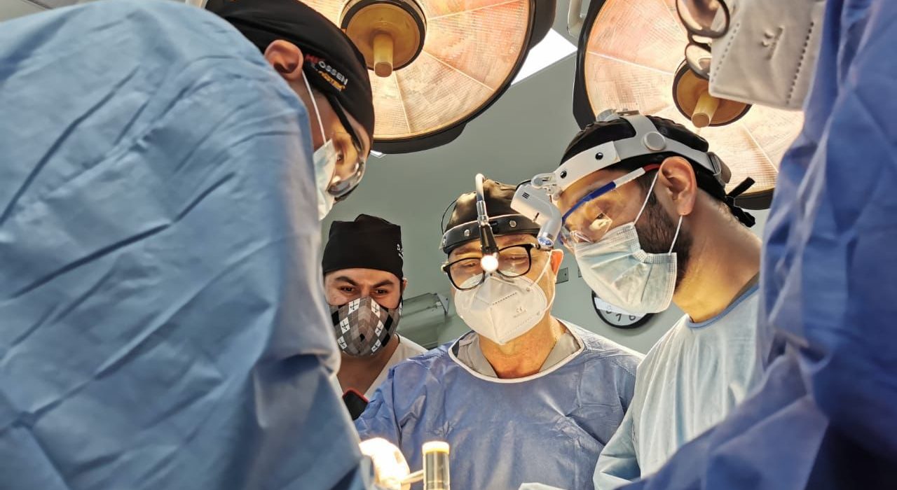
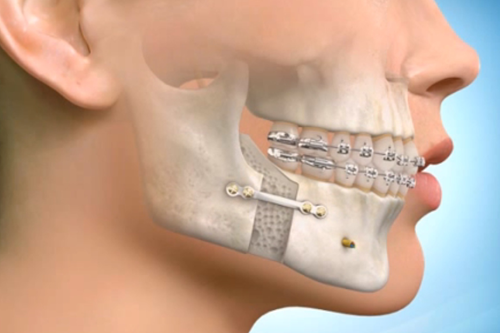
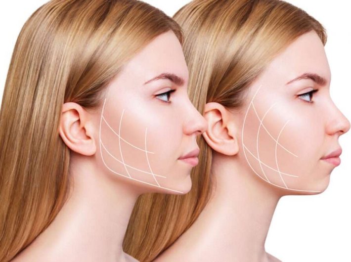
Orthognathic surgery, also known as corrective jaw surgery is the use of surgical procedures to correct imbalance in the upper and lower jaws. These procedures may be used to treat an abnormality where the teeth do not fit together properly (malocclusion), to treat airway obstruction when a patient is asleep (obstructive sleep apnea), or to improve the balance and overall appearance of the face. Often patients who are undergoing orthodontic treatment with braces may find that an imbalance of their jaws make it impossible for their bite to be corrected without repositioning the jaws. Treatment with orthognathic surgery is carried out with combined management between an orthodontist and a maxillofacial surgeon.

What Kinds of Conditions are treated with Orthognathic Surgery?

Around 20% of patients have dental and jaw abnormalities that cannot be corrected with braces alone and may be candidates for orthognathic surgery. Patients undergoing braces with an orthodontist may wish to discuss if they will require orthognathic surgery as part of their treatment.
Additionally, many craniofacial syndromes and conditions such as cleft lip and cleft palate, syndromic craniosynostosis (Apert, Crouzon, Pfeiffer, Muenke, Saethre-Chotzen, etc.), Hemifacial microsomia, Treacher Collins syndrome, Miller syndrome and Nager syndrome cause imbalances of the upper and lower jaws that are improved with orthognathic surgery.

Benefits of Orthognathic Surgery?

Patients may suffer significant wear and injury to their teeth over time if they have a condition where their teeth do not fit together properly (malocclusion). In cases with significant malocclusion, surgery may been needed to place the jaws in the correct position in order to achieve a normal bite that will improve the appearance and life span of the teeth.
Patients who have significant trouble breathing at night (obstructive sleep apnea) may benefit from having their jaws advanced to open their airway in the back of their throat. Patients often have significant improvement or are cured from their sleep apnea when these procedure are performed in the correct patient.
Along with the functional concerns related to a patient’s teeth and breathing, orthognathic operations may dramatically enhance a patient’s appearance. Not all patients experience the same benefit, but correction of significant jaw discrepancies may result in a more harmonious facial balance, and expansion of the facial skeleton often results in changes that make a patient appear youthful for longer.

How are Patients Evaluated for Orthognathic Surgery?

Orthognathic surgery is completed with a team comprised of a maxillofacial surgeon and an orthodontist. The orthodontist uses braces and other appliances to align the teeth appropriately in the upper jaw (maxilla) and lower jaw (mandible). Prior to beginning treatment, the orthodontists will take a series of X-rays, photographs, and dental impressions.
Extensive analysis of these will be used to determine the best treatment plan to align your teeth and improve your appearance. At this time, your orthodontist may discuss with you if they think jaw surgery will be needed to address all of your concerns. Once the orthodontist is nearing completion of your treatment, a full set of orthodontic records will be taken again so we can plan your operation. This includes X-rays, photographs, and dental impressions.

Facial fractures (facial trauma)

A facial fracture is a broken bone in the face. The face has a complex bone structure. The facial skeleton consists of the:
  • Frontal bone (forehead).
  • Zygomas (cheekbones).
  • Orbital bones (eye sockets).
  • Nasal bones.
  • Maxillary bones (upper jaw).
  • Mandible (lower jaw).

There are many other bones that are found deeper within the facial structure. Muscles required for chewing, swallowing and talking are attached to these bones.

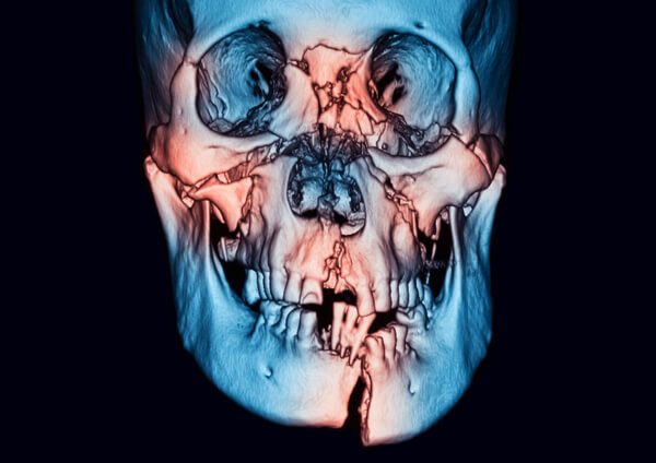
Nasal fractures (broken nose) are the most common. Fractures to other facial bones can also occur. You might only have one fracture, or you might have several broken bones. Multiple fractures are more likely to occur during a motor vehicle accident or other high-impact accident. Fractures may be unilateral (occurring on one side of the face) or bilateral (occurring on both sides of the face).

Is a facial fracture a serious problem?

If you suffer from a facial injury, you should seek immediate medical attention. Some fractures are minor. However, complex fractures may cause irreversible damage and can even be life-threatening.

Located near to the bones in your face are the nerves and muscles that are responsible for sensations, expressions and eye movements. The muscles and nerves are located near to the facial bones. The face is close to the brain and central nervous system (CNS). Fractures may result in damage to cranial nerves, depending on the particular type and location of the fracture. Fractures to the orbit (eye socket) may result in problems with vision. Fractures of the nose may make it difficult for the injured person to breathe or smell. Also, fractures of the jawbones may cause breathing problems or make it difficult to chew, speak, or swallow.

What are the kinds of facial fractures?

There are several main types of facial fractures.

Nasal bones (broken nose): Nasal bone fractures are the most common type of facial fracture. The nasal bone is made up of two thin bones. It takes less force to break the nasal bones than other facial bones because they are thin and prominent. Usually, the nose looks deformed or feels sore to the touch after a fracture. Swelling in the area might make it more difficult to assess how much damage has occurred. Nosebleeds and bruising around the nose are common symptoms of a nasal fracture.

Symptoms of a nose fracture may include:

  • Purplish patch on skin caused when blood leaks from broken blood vessels (also called bruising or ecchymosis).
  • Discoloration under the eyes (“black eyes”).
  • Blockage of one or both nostrils or a deviated septum.
  • Twisted or crooked nose or indented bridge.
  • Nosebleed.Frontal bone (forehead) fractures: The frontal bone is the main bone in the forehead area. A high-impact injury to the head can cause a fracture of the frontal bone and floor of the sinuses. The fracture is mostly likely to occur in the middle of the forehead. That’s where the bone is the thinnest and weakest. An injury may cause the bone to be indented (pushed inward). Substantial force is required to fracture the frontal bone, so often other injuries to the face and skull or neurological trauma may be present. Associated problems may include leakage of the cerebrospinal fluid, eye injuries and damage to the sinus ducts.
  • Zygomaticomaxillary fractures (broken cheekbone/upper jaw): The zygomas (cheekbones) are attached at several points to the upper jaw (maxilla) and bones of the skull. Fractures to the cheekbone(s) might also involve breaks in other facial bones nearby.
  • Orbital fractures (eye socket): There are three main types of orbital fractures.
  • Orbital rim fracture: The outer rim is the thickest part of the eye socket. It requires a lot of force to break the bone. Many other injuries may accompany an orbital rim fracture, such as damage to the optic nerve.
  • Blowout fractures: The orbital rim remains intact in this case, but a crack forms in the thin bone at the lower part of the eye socket. The eye muscles and other structures can become entrapped in the break and prevent the eyeball from moving normally.
  • Direct orbital floor fracture: This is a rim fracture that extends into the lower socket.
Symptoms of an orbital fracture may include:
  • Blurry, decreased or double vision (diplopia).
  • Difficulty in moving eyes left, right, up or down.
  • Swollen forehead or cheek or swelling under the eyes.
  • Flatness of the cheeks.
    Sunken or bulging eyeballs.
  • Facial numbness near the injury.
  • Blood or discoloration in the white part of the eye.

Mid-face (Le Fort fractures): Blunt force trauma tends to cause fractures along three lines of weakness in the mid-face. One characteristic of all types of Le Fort fractures is the fracture of the pterygoid processes, part of the sphenoid bone. There are three main types of Le Fort fractures, but there may be individual variations.

  • Le Fort I: The fracture extends above the upper jaw (maxilla).
  • Le Fort II: The fracture extends from the lower part of one cheek, below the eye, across the bridge of the nose, and to the lower part of the other cheek.
  • Le Fort III: The fracture extends across the bridge of the nose and the bones surrounding the eyes.

Mandible (lower jaw): The mandible holds the lower teeth in place and moves when you are talking or chewing. Fractures of the lower jaw affect the sections of the lower jaw that supports teeth (called the body), the part where the jaw curves upwards into the neck (the angle) or the knob-shaped joint at the top of the jaw bone (the condyle) or the point where the two sides of the lower jaw are joined (the symphysis). If you have a break in the lower jaw, you may also have broken or loose teeth.

Symptoms of upper or lower jaw fractures:
  • Trouble with chewing, eating, or speaking.
  • Loose, broken or missing teeth.
  • Teeth not fitting together properly.
  • Cheek pain when opening the mouth.

What causes facial fractures?

You can break the bones in your face in many ways, including:
  • High-impact accidents, such as motor vehicle accidents.
  • Sports injuries.
  • Workplace accidents.
    Falls.
  • Interpersonal trauma like fighting or domestic violence.

MANAGEMENT AND TREATMENT

How are facial fractures treated?

Your healthcare provider may prescribe pain-relieving drugs as well as oral steroids to ease swelling. Your provider will prescribe antibiotics if there’s a high risk of infection.

In general, fractures may be treated by performing a closed reduction (resetting the broken bone or bones without surgery) or an open reduction (surgery that requires an incision to reposition the fractured bones). For a complex fracture with multiple broken bones, you will need reconstructive surgery.

The type of treatment will depend on the location and extent of the injury. The aim of treatment for facial fractures is to restore the normal appearance and function of the injured areas.

Buccal Fat Pad Removal

Feeling confident and good about yourself is important. Although some fullness in the cheeks can offer a youthful appearance, too much can make you feel ‘chubby-cheeked’. This can make you self-conscious about the way you look. Buccal fat removal is the surgical process of removing fat from the cheeks. The safe and effective procedure can accentuate the contour of the cheeks and create a more defined and balanced facial appearance.

What is Buccal Fat Pad Removal?

Buccal fat removal is also known as buccal fat pad excision and cheek reduction surgery. The procedure targets your buccal fat pad which is the rounded fat in the middle of the cheek. It’s in the hollow area beneath your cheekbone, between your facial muscles. Everyone has buccal fat pads, but the size can vary from one person to another.

What Happens During Fat Pad Removal Surgery?

The surgery is performed from inside the mouth. This means that there are no visible scars. The procedure is relatively simple and straightforward. The procedure takes around 40 minutes.
Your maxillofacial surgeon makes a small incision inside the cheek under local anaesthetic. The incisions are about 5 millimeters long. The buccal fat pad is located between each cheekbone and the buccinator muscle (the muscle that helps with chewing). Your surgeon will then remove the buccal fat pad. Excess fat is then removed from the cheeks using a fine-tipped surgical tool to create a more defined and angular face. The incision is then closed using dissolvable sutures.
Typically, patients find that buccal fat pad removal can dramatically affect their entire facial structure, cheekbones, eyes and jawline. In a suitable candidate, patients find that the procedure results in a slimmer midface, similar to sucking in your cheeks.

How Long Does it Take to Recover from Buccal Fat Pad Removal?

Buccal fat pad removal is growing in popularity for two main reasons: minimal downtime and no visible scarring. You can return to daily activities within roughly 7 days of the surgery. Make sure to pencil in some downtime after your surgery to fully rest and recover. You should see the final results in 3- 4 weeks once all the swelling has gone down.

What Are the Benefits of Buccal Fat Removal?

After undergoing surgery, patients can expect a slimmer, more contoured face. The procedure not only affects how your cheeks look but the overall appearance of your face. Buccal fat removal has several benefits, including:
  • Contour cheekbones
  • Reduce fat volume in cheeks
  • More chiselled jawline and cheeks

How Long Will Results Last?

The results from the procedure are permanent. Once you remove those fat pads, they are gone for good. Although your buccal fat pads won’t grow back, weight gain can mask the results of the procedure. As you age, you may see fat start to descend into the lower part of the face as well.

Your face will likely look fuller during your recovery. But after the swelling goes down, you should start to see the final result. Swelling should subside in approximately 3 weeks. It can take as long as 3 to 6 months to see the final result. During this time, your skin is settling into its new position after the fat is hollowed out.

Is Buccal Fat Pad Removal Painful?

Most patients don’t experience pain during the procedure because of the local anaesthetic. There may be some discomfort in recovery. Your mouth and jawline may feel sore, sensitive, heavy, or hard following your surgery. This should subside during recovery as you heal.

Bone Grafting

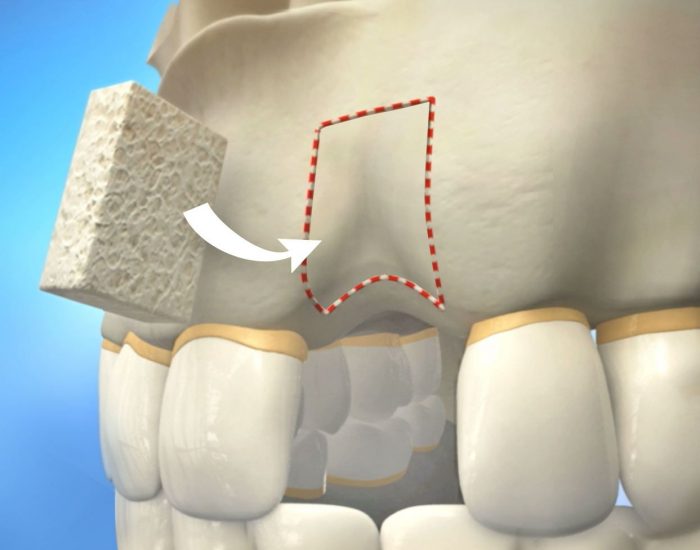
Over a period of time, the jawbone associated with missing teeth atrophies (shrinks) or recedes. This often results in the presence of less than adequate bone density/volume suitable for placement of dental implants. In the past, most patients in this situation were denied the placement of dental implants.

Today, we have the techniques and biomaterials to graft or grow bone where needed. This not only gives us the opportunity to place implants of sufficient sizes in the prosthodontics guided 3D position, it also gives us a chance to restore functionality and aesthetic appearance.

MINOR GRAFTS OR AUGMENTATIONS

Minor bone grafting such as immediate socket augmentation after an extraction, can repair potential implant sites with damaged bone structure due to previous extractions, infections, gum disease or injuries.

There are different Biomaterials that are commonly used for minor augmentations with “bone in a bottle”. These have animal, human or purely synthetic origin and include: a) bone elements of cow bone origin (xenograft), b) bone from human donor origin obtained by tissue banks as part of multi-organ donor scheme, c) synthetic bone of calcium phosphate origin or d) your own bone is taken from the jaw, hip or tibia (below the knee). For minor augmentations normally xenografts of cow bone origin are used with long term safety and efficacy shown through extensive clinical studies spanning over decades both in orthopaedic surgery and oral surgery.

We use your own growth factors obtained from a sample of your own blood to accelerate and promote bone and soft tissue formation in graft areas. This regenerative medicine technique is known as Platelet Rich Plasma (PRP) or Platelet Rich Fibrin (PRF).

Socket Preservation Bone Graft

What is Tooth Socket Preservation Bone Graft?

A socket preservation graft, also known as “alveolar ridge preservation graft”, is a surgical procedure used to fill in a socket after tooth extraction. At the time of tooth removal, an oral & maxillofacial surgeon will place special bone grafting material in the extraction socket to avoid post-extraction bone loss and accommodate future replacement tooth options like dental implants or dental bridges.
Placing an extraction socket preservation graft at the time of tooth extraction doesn’t require a separate surgery and doesn’t cause any additional discomfort, as there is already exposed bone post – extraction.

Is Bone Graft Necessary after Tooth Extraction?

No, socket preservation bone grafts aren’t necessarily required every time a tooth is extracted, – this depends on the patient’s age, biotype, the location of the extracted tooth, and the overall oral health of the patient. In some cases, extraction sites can heal completely fine on their own, resulting in sufficient bony ridge height and horizontal ridge width for implant placement.

However, with complicated extractions, a lot of alveolar bone can be lost, and adding a bone graft at the time of removal can prevent the socket walls from caving in, providing a solid, firm foundation for the future dental implant.

Benefits of Socket Preservation Grafting

Socket grafting is often used if the patient decides to proceed with restorative procedures which not only include dental implants but also dental bridges and dentures.
  • Implants. A dental implant candidate must have a minimum amount of jaw bone to surround the implant in order to establish long-term stability and success. Without adequate bone volume, the patient may experience accelerated bone and soft tissue loss around the implant over time. A socket preservation graft is used to ensure that you will have enough bone to enjoy your dental implants for a lifetime.
  • Dental bridges A graft after a tooth extraction will help to preserve proper bone height to prevent bone loss and lowering gum tissue levels. Without a socket preservation graft, you are likely to see a growing space between an opposing bridge and recessed gum. To improve the aesthetics of the future dental bridge, the bone grafting material will be applied at the time of extraction.
  • Dentures. Most dentists choose not to conduct ridge preservation procedures and extraction site reconstruction before denture therapy. However, the stability, fit, and retention of dentures are improved with a higher quantity of jawbone. As such, your dentist may recommend placing socket preservation grafts in strategic areas after extractions.
  • No post-extraction tooth replacement. Even if patients choose not to replace the lost tooth, an alveolar ridge preservation graft can still have its benefits. Gum recession following alveolar ridge resorption at the extraction site could expose roots of the neighboring teeth, leading to increased sensitivity.

RIDGE AUGMENTATION:

The jawbone will lose 30-40% of its volume within the first 3 months after tooth extraction. Bone loss can result in oral health issues and physical changes to your facial profile. This could cause significant changes to your overall dental function, phonetics of your speech and aesthetic appearance of your face. Jaw bone Ridge augmentation is a reconstructive method for replacing the missing bone and gum tissue to restore proper anatomy, contour and function, as part of the dental implant process. The ridge augmentation process involves sculpturing the natural contour of the jaw and gums using bone and/or gum graft material. Once the area has healed and the contour of the alveolar ridge is restored, the dental implants are placed at a later stage.

MAJOR GRAFTS OR AUGMENTATIONS

Major bone grafts are typically performed to repair moderate to large amounts of bone loss or defects of the jaws. These defects may arise as a result of traumatic injuries, tumor surgery, or congenital defects. Very large defects (e.g. after extensive cancer surgery or road traffic accidents) are repaired typically using the patient’s own bone. This bone is harvested from a number of different sites depending on the size of the defect. The lower or upper jaw, the hip (iliac crest), and lateral knee (tibia), are common donor sites. These procedures are routinely performed in an operating room and may require a hospital stay. Fortunately, these procedures are exceptional and infrequently required for the majority of patients.

SINUS LIFT BONE GRAFTS

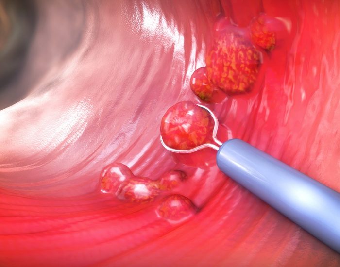
Sinus lift bone grafts are indicated commonly to replace bone in the back of the upper jaw when replacing back teeth (molars).

The Maxillary sinus is a medium size empty air cavity located within the upper jaw bone where the roots of molar teeth are normally embedded. When these upper teeth are removed, there can be a thin layer of bone left to separate the maxillary sinus from the mouth. When the sinus wall is very thin, it may not be possible to place traditional dental implants in this area. The procedure to augment or grow bone below the floor of the maxillary sinus cavity is called a “sinus lift graft” or “sinus augmentation”.

To place implants, the floor of this air cavity bordering the teeth is filled with bone substitutes or biomaterials. A xenograft (cow bone origin) is the material of choice for this procedure used very safely and successfully over decades. Special dissolving membranes are used under the gum to protect the bone graft and encourage bone regeneration. This is called guided bone regeneration (GBR). Membranes can be synthetic or porcine in origin.

If there is a borderline amount of bone remaining between the ridge of the upper jaw and the floor of the sinus cavity to stabilize the implant, sinus augmentations and dental implant placement can sometimes be performed as a single procedure. This is known as “simultaneous sinus lift” meaning that it is carried out at the time of implant placement. If not enough bone is available, the sinus lift is performed first and the graft is allowed to mature for a few months ( 6 to 8 months). Once the graft has matured, the implants can be placed. This is referred to as “staged sinus lift”.

Oral pathology & Biopsies

Oral Pathology refers to all diseases of the oral region, including cysts, tumors, infections, and others. Many oral pathologies are simple to treat, but some can cause serious complications if they go unnoticed. Our oral & maxillofacial surgeon, recommends regular oral examinations as the best way to prevent and control oral pathologies. If you notice any irregularities in the lining of your mouth, make an appointment to have your mouth examined by an expert. Oral & maxillofacial surgeons undergo extensive training to diagnose and treat all sorts of oral pathologies, and we will gladly schedule your appointment as soon as possible.